Pages

Wednesday, May 19, 2010

CANCER LINKED TO MISSING DNA


Scientists reported the first link ever found between cancer and a type of genetic defect, called copy number variation, characterized by missing or extra bits of DNA.

The breakthrough came in a study on neuroblastoma, a devastating pediatric disease of the nervous system that accounts for 15 percent of all cancer deaths among young children.

Researchers led by John Maris of Children's Hospital of Philadelphia discovered that a copy number variation (CNV) on chromosome 1 can play a key role in the development of the disease, which strikes most commonly in infancy and is often fatal.

The absent stretch of DNA occurs within a group of genes involved in the development of the nervous system, and affects how much one of these genes is manufactured within both normal and the cancerous cells.

"This is a brand new area -- we never suspected that this family of genes played a role in neuroblastoma," Maris said in an phone interview.

Tuesday, May 18, 2010

NEW WAY TO STORE INFORMATION VIA DNA-DISCOVERED


Researchers at UC Riverside have found a way to get into your body and your bloodstream. No, they’re not spiritual gurus or B-movie mad scientists. Nathaniel G. Portney, Yonghui Wu, Stefano Lonardi, and Mihri Ozkan from UCR’s departments of Bioengineering, Computer Science and Engineering, Biochemistry, and Electrical Engineering, and the Center for Nanoscale Science and Engineering, are just talented when it comes to manipulating DNA.

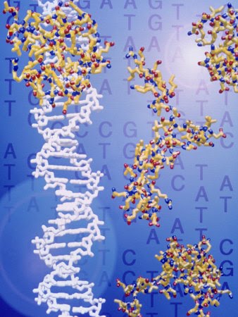
The researchers discovered a system to encode digital information within DNA. This method relies on the length of the fragments obtained by the partial restriction digest rather than the actual content of the nucleotide sequence. As a result, the technology eliminates the need to use expensive sequencing machinery.

Why is this discovery important? The human genome consists of the equivalent of approximately 750 megabytes of data – a significant amount of storage space. However, only about three percent of DNA goes into composing the more than 22,000 genes that make us what we are. The remaining 97 percent leaves plenty of room to encode information in a genome, allowing the information to be preserved and replicated in perpetuity.

Given the size of the DNA fragments (one base pair of DNA is 0.33 nanometers), one could store a large amount of information in a very small space. By storing messages within DNA, organizations can “tag” objects to verify authenticity, as well as to inconspicuously send data to a specific destination. “Already there are several companies using DNA to tag objects that they certify to be original and which then can be very difficult to counterfeit,” says Stefano Lonardi, Associate Professor of Computer Science & Engineering at UCR’s Bourns College of Engineering.

For example, the British company, Redweb Security, has developed something called i-powder that tags DNA and another company called PSA DNA Authentication services tags sports memorabilia.

“What we developed at UCR is a method to encode a message in DNA in a way that does not require an expensive sequencing machine,” notes Lonardi. “The decoding still requires a wet lab procedure, but the experimental procedure is significantly easier.”

The article, entitled “Length-based Encoding of Binary Data in DNA,” was published by the American Chemical Society in Langmuir December 18, 2007.

GENE THERAPHY


Genes, which are carried on chromosomes, are the basic physical and functional units of heredity. Genes are specific sequences of bases that encode instructions on how to make proteins. Although genes get a lot of attention, it’s the proteins that perform most life functions and even make up the majority of cellular structures. When genes are altered so that the encoded proteins are unable to carry out their normal functions, genetic disorders can result.

Gene therapy is a technique for correcting defective genes responsible for disease development. Researchers may use one of several approaches for correcting faulty genes:

normal gene may be inserted into a nonspecific location within the genome to replace a nonfunctional gene. This approach is most common.

abnormal gene could be swapped for a normal gene through homologous recombination.

abnormal gene could be repaired through selective reverse mutation, which returns the gene to its normal function.

regulation (the degree to which a gene is turned on or off) of a particular gene could be altered.

Monday, May 17, 2010

GENES AND GENOMES


Genomic DNA is located in the cell nucleus of eukaryotes, as well as small amounts in mitochondria and chloroplasts. In prokaryotes, the DNA is held within an irregularly shaped body in the cytoplasm called the nucleoid. The genetic information in a genome is held within genes, and the complete set of this information in an organism is called its genotype. A gene is a unit of heredity and is a region of DNA that influences a particular characteristic in an organism. Genes contain an open reading frame that can be transcribed, as well as regulatory sequences such as promoters and enhancers, which control the transcription of the open reading frame.

In many species, only a small fraction of the total sequence of the genome encodes protein. For example, only about 1.5% of the human genome consists of protein-coding exons, with over 50% of human DNA consisting of non-coding repetitive sequences. The reasons for the presence of so much non-coding DNA in eukaryotic genomes and the extraordinary differences in genome size, or C-value, among species represent a long-standing puzzle known as the "C-value enigma."However, DNA sequences that do not code protein may still encode functional non-coding RNA molecules, which are involved in the regulation of gene expression.

Some non-coding DNA sequences play structural roles in chromosomes. Telomeres and centromeres typically contain few genes, but are important for the function and stability of chromosomes. An abundant form of non-coding DNA in humans are pseudogenes, which are copies of genes that have been disabled by mutation. These sequences are usually just molecular fossils, although they can occasionally serve as raw genetic material for the creation of new genes through the process of gene duplication and divergence.

QUADRUPLUX STRUCTURE OF DNA


At the ends of the linear chromosomes are specialized regions of DNA called telomeres. The main function of these regions is to allow the cell to replicate chromosome ends using the enzyme telomerase, as the enzymes that normally replicate DNA cannot copy the extreme 3' ends of chromosomes. These specialized chromosome caps also help protect the DNA ends, and stop the DNA repair systems in the cell from treating them as damage to be corrected. In human cells, telomeres are usually lengths of single-stranded DNA containing several thousand repeats of a simple TTAGGG sequence.

These guanine-rich sequences may stabilize chromosome ends by forming structures of stacked sets of four-base units, rather than the usual base pairs found in other DNA molecules. Here, four guanine bases form a flat plate and these flat four-base units then stack on top of each other, to form a stable G-quadruplex structure. These structures are stabilized by hydrogen bonding between the edges of the bases and chelation of a metal ion in the centre of each four-base unit. Other structures can also be formed, with the central set of four bases coming from either a single strand folded around the bases, or several different parallel strands, each contributing one base to the central structure.

In addition to these stacked structures, telomeres also form large loop structures called telomere loops, or T-loops. Here, the single-stranded DNA curls around in a long circle stabilized by telomere-binding proteins. At the very end of the T-loop, the single-stranded telomere DNA is held onto a region of double-stranded DNA by the telomere strand disrupting the double-helical DNA and base pairing to one of the two strands. This triple-stranded structure is called a displacement loop or D-loop.

ALTERNATE STRUCTURE OF DNA


DNA exists in many possible conformations that include A-DNA, B-DNA, and Z-DNA forms, although, only B-DNA and Z-DNA have been directly observed in functional organisms. The conformation that DNA adopts depends on the hydration level, DNA sequence, the amount and direction of supercoiling, chemical modifications of the bases, the type and concentration of metal ions, as well as the presence of polyamines in solution.
The first published reports of A-DNA X-ray diffraction patterns— and also B-DNA used analyses based on Patterson transforms that provided only a limited amount of structural information for oriented fibers of DNA. An alternate analysis was then proposed by Wilkins et al., in 1953, for the in vivo B-DNA X-ray diffraction/scattering patterns of highly hydrated DNA fibers in terms of squares of Bessel functions. In the same journal, Watson and Crick presented their molecular modeling analysis of the DNA X-ray diffraction patterns to suggest that the structure was a double-helix.

Although the `B-DNA form' is most common under the conditions found in cells, it is not a well-defined conformation but a family of related DNA conformations that occur at the high hydration levels present in living cells. Their corresponding X-ray diffraction and scattering patterns are characteristic of molecular paracrystals with a significant degree of disorder.

Compared to B-DNA, the A-DNA form is a wider right-handed spiral, with a shallow, wide minor groove and a narrower, deeper major groove. The A form occurs under non-physiological conditions in partially dehydrated samples of DNA, while in the cell it may be produced in hybrid pairings of DNA and RNA strands, as well as in enzyme-DNA complexes.Segments of DNA where the bases have been chemically modified by methylation may undergo a larger change in conformation and adopt the Z form. Here, the strands turn about the helical axis in a left-handed spiral, the opposite of the more common B form.These unusual structures can be recognized by specific Z-DNA binding proteins and may be involved in the regulation of transcription.

Sunday, May 16, 2010

DNA SEQUENCE


The primary structure of a biological molecule is the exact specification of its atomic composition and the chemical bonds connecting those atoms. As nucleic acids, e.g. DNA and RNA, are unbranched polymers, this is equivalent to specifying exact sequence of nucleotides that comprise the whole molecule. This sequence is written as a succession of letters representing a real or hypothetical DNA molecule or strand. By convention, the primary structure of a DNA or RNA molecule is reported from the 5' end to the 3' end.
The sequence has capacity to carry information. When used in reference to biological DNA, which carries the information which directs the functions of living beings, the term genetic sequence is often used. Sequences can be read from the biological raw material through DNA sequencing methods.
Primary structure is sometimes mistakenly termed primary sequence, but there is no such term, as well as no parallel concept of secondary or tertiary sequence.

Notation :

Main article: Nucleic acid notation
While A, T, C, and G represent a particular nucleotide at a position, there are also letters that represent ambiguity. Of all the molecules sampled, there is more than one kind of nucleotide at that position. The rules of the International Union of Pure and Applied Chemistry (IUPAC) are as follows:[1]
A = adenine
C = cytosine
G = guanine
T = thymine
R = G A (purine)
Y = T C (pyrimidine)
K = G T (keto)
M = A C (amino)
S = G C (strong bonds)
W = A T (weak bonds)
B = G T C (all but A)
D = G A T (all but C)
H = A C T (all but G)
V = G C A (all but T)
N = A G C T (any)
These symbols are also valid for RNA, although U is often written instead of T (for uracil rather than thymine)

DNA REPAIR


DNA repair refers to a collection of processes by which a cell identifies and corrects damage to the DNA molecules that encode its genome. In human cells, both normal metabolic activities and environmental factors such as UV light and radiation can cause DNA damage, resulting in as many as 1 million individual molecular lesions per cell per day.Many of these lesions cause structural damage to the DNA molecule and can alter or eliminate the cell's ability to transcribe the gene that the affected DNA encodes. Other lesions induce potentially harmful mutations in the cell's genome, which affect the survival of its daughter cells after it undergoes mitosis. Consequently, the DNA repair process is constantly active as it responds to damage in the DNA structure. When normal repair processes fail, and when cellular apoptosis does not occur, irreparable DNA damage may occur, including double-strand breaks and DNA crosslinkages.

The rate of DNA repair is dependent on many factors, including the cell type, the age of the cell, and the extracellular environment. A cell that has accumulated a large amount of DNA damage, or one that no longer effectively repairs damage incurred to its DNA, can enter one of three possible states:

1. A irreversible state of dormancy, known as senescence
2. Cell suicide, also known as apoptosis or programmed cell death
3. Unregulated cell division, which can lead to the formation of a tumor that is cancerous

The DNA repair ability of a cell is vital to the integrity of its genome and thus to its normal functioning and that of the organism. Many genes that were initially shown to influence life span have turned out to be involved in DNA damage repair and protection.Failure to correct molecular lesions in cells that form gametes can introduce mutations into the genomes of the offspring and thus influence the rate of evolution.

DNA


Deoxyribonucleic acid (DNA) is a nucleic acid that contains the genetic instructions used in the development and functioning of all known living organisms and some viruses. The main role of DNA molecules is the long-term storage of information. DNA is often compared to a set of blueprints or a recipe, or a code, since it contains the instructions needed to construct other components of cells, such as proteins and RNA molecules. The DNA segments that carry this genetic information are called genes, but other DNA sequences have structural purposes, or are involved in regulating the use of this genetic information.

Chemically, DNA consists of two long polymers of simple units called nucleotides, with backbones made of sugars and phosphate groups joined by ester bonds. These two strands run in opposite directions to each other and are therefore anti-parallel. Attached to each sugar is one of four types of molecules called bases. It is the sequence of these four bases along the backbone that encodes information. This information is read using the genetic code, which specifies the sequence of the amino acids within proteins. The code is read by copying stretches of DNA into the related nucleic acid RNA, in a process called transcription.

Within cells, DNA is organized into long structures called chromosomes. These chromosomes are duplicated before cells divide, in a process called DNA replication. Eukaryotic organisms (animals, plants, fungi, and protists) store most of their DNA inside the cell nucleus and some of their DNA in organelles, such as mitochondria or chloroplasts. In contrast, prokaryotes (bacteria and archaea) store their DNA only in the cytoplasm. Within the chromosomes, chromatin proteins such as histones compact and organize DNA. These compact structures guide the interactions between DNA and other proteins, helping control which parts of the DNA are transcribed.

GENETIC ENGINEERING


Genetic engineering, recombinant DNA technology, genetic modification/manipulation , and gene splicing are terms that apply to the direct manipulation of an organism's genes.Genetic engineering is different from traditional breeding, where the organism's genes are manipulated indirectly. Genetic engineering uses the techniques of molecular cloning and transformation to alter the structure and characteristics of genes directly. Genetic engineering techniques have been applied to various industries, principally medicine and agriculture, with some success. Examples include production of synthetic human insulin using modified bacteria, production of erythropoietin in hamster ovary cells, the production of new types of experimental mice such as the oncomouse (cancer mouse) for research purposes, and the production of new strains of crop plants with beneficial qualities.

The term "genetic engineering" was coined by Jack Williamson in his science fiction novel Dragon's Island, published in 1951, one year before DNA's role in heredity was confirmed by Alfred Hershey and Martha Chase, and two years before James Watson and Francis Crick showed that the DNA molecule has a double-helix structure.

DNA PROFILING


DNA profiling is a technique employed by forensic scientists to assist in the identification of individuals on the basis of their respective DNA profiles. DNA profiles are encrypted sets of numbers that reflect a person's DNA makeup, which can also be used as the person's identifier. DNA profiling should not be confused with full genome sequencing. It is used in, for example, parental testing and rape investigation.

Although 99.9% of human DNA sequences are the same in every person, enough of the DNA is different to distinguish one individual from another.DNA profiling uses repetitive sequences that are highly variable,called variable number tandem repeats (VNTR). VNTRs loci are very similar between closely related humans, but so variable that unrelated individuals are extremely unlikely to have the same VNTRs.

The DNA profiling technique was first reported in 1984 by Sir Alec Jeffreys at the University of Leicester in England,and is now the basis of several national DNA databases. Dr. Alec Jeffrey's genetic fingerprinting was made commercially available in 1987, when a chemical company, ICI, started a blood-testing center in England.

GENETIC CODE


Genes generally express their functional effect through the production of proteins, which are complex molecules responsible for most functions in the cell. Proteins are chains of amino acids, and the DNA sequence of a gene is used to produce a specific protein sequence. This process begins with the production of an RNA molecule with a sequence matching the gene's DNA sequence, a process called transcription.

This messenger RNA molecule is then used to produce a corresponding amino acid sequence through a process called translation. Each group of three nucleotides in the sequence, called a codon, corresponds to one of the twenty possible amino acids in protein – this correspondence is called the genetic code. The flow of information is unidirectional: information is transferred from nucleotide sequences into the amino acid sequence of proteins, but it never transfers from protein back into the sequence of DNA—a phenomenon Francis Crick called the central dogma of molecular biology.
The dynamic structure of hemoglobin is responsible for its ability to transport oxygen within mammalian blood.
A single amino acid change causes hemoglobin to form fibers.

The specific sequence of amino acids results in a unique three-dimensional structure for that protein, and the three-dimensional structures of protein are related to their function.Some are simple structural molecules, like the fibers formed by the protein collagen. Proteins can bind to other proteins and simple molecules, sometimes acting as enzymes by facilitating chemical reactions within the bound molecules (without changing the structure of the protein itself). Protein structure is dynamic; the protein hemoglobin bends into slightly different forms as it facilitates the capture, transport, and release of oxygen molecules within mammalian blood.

A single nucleotide difference within DNA can cause a single change in the amino acid sequence of a protein. Because protein structures are the result of their amino acid sequences, some changes can dramatically change the properties of a protein by destabilizing the structure or changing the surface of the protein in a way that changes its interaction with other proteins and molecules. For example, sickle-cell anemia is a human genetic disease that results from a single base difference within the coding region for the ß-globin section of hemoglobin, causing a single amino acid change that changes hemoglobin's physical properties.Sickle-cell versions of hemoglobin stick to themselves, stacking to form fibers that distort the shape of red blood cells carrying the protein. These sickle-shaped cells no longer flow smoothly through blood vessels, having a tendency to clog or degrade, causing the medical problems associated with this disease.

Some genes are transcribed into RNA but are not translated into protein products—these are called non-coding RNA molecules. In some cases, these products fold into structures which are involved in critical cell functions .RNA can also have regulatory effect through hybridization interactions with other RNA molecules

MOLECULAR GENETICS

Although genes were known to exist on chromosomes, chromosomes are composed of both protein and DNA—scientists did not know which of these is responsible for inheritance. In 1928, Frederick Griffith discovered the phenomenon of transformation dead bacteria could transfer genetic material to "transform" other still-living bacteria. Sixteen years later, in 1944, Oswald Theodore Avery, Colin McLeod and Maclyn McCarty identified the molecule responsible for transformation as DNA.The Hershey-Chase experiment in 1952 also showed that DNA (rather than protein) is the genetic material of the viruses that infect bacteria, providing further evidence that DNA is the molecule responsible for inheritance.

James D. Watson and Francis Crick determined the structure of DNA in 1953, using the X-ray crystallography work of Rosalind Franklin and Maurice Wilkins that indicated DNA had a helical structure (i.e., shaped like a corkscrew). Their double-helix model had two strands of DNA with the nucleotides pointing inward, each matching a complementary nucleotide on the other strand to form what looks like rungs on a twisted ladder.This structure showed that genetic information exists in the sequence of nucleotides on each strand of DNA. The structure also suggested a simple method for duplication: if the strands are separated, new partner strands can be reconstructed for each based on the sequence of the old strand.

Although the structure of DNA showed how inheritance works, it was still not known how DNA influences the behavior of cells. In the following years, scientists tried to understand how DNA controls the process of protein production. It was discovered that the cell uses DNA as a template to create matching messenger RNA (a molecule with nucleotides, very similar to DNA). The nucleotide sequence of a messenger RNA is used to create an amino acid sequence in protein; this translation between nucleotide and amino acid sequences is known as the genetic code.

With this molecular understanding of inheritance, an explosion of research became possible. One important development was chain-termination DNA sequencing in 1977 by Frederick Sanger: This technology allows scientists to read the nucleotide sequence of a DNA molecule. In 1983, Kary Banks Mullis developed the polymerase chain reaction, providing a quick way to isolate and amplify a specific section of a DNA from a mixture.Through the pooled efforts of the Human Genome Project and the parallel private effort by Celera Genomics, these and other techniques culminated in the sequencing of the human genome in 2003.

GENETICS


The fact that living things inherit traits from their parents has been used since prehistoric times to improve crop plants and animals through selective breeding. However, the modern science of genetics, which seeks to understand the process of inheritance, only began with the work of Gregor Mendel in the mid-nineteenth century. Although he did not know the physical basis for heredity, Mendel observed that organisms inherit traits via discrete units of inheritance, which are now called genes.

Genes correspond to regions within DNA, a molecule composed of a chain of four different types of nucleotides—the sequence of these nucleotides is the genetic information organisms inherit. DNA naturally occurs in a double stranded form, with nucleotides on each strand complementary to each other. Each strand can act as a template for creating a new partner strand—this is the physical method for making copies of genes that can be inherited.

The sequence of nucleotides in a gene is translated by cells to produce a chain of amino acids, creating proteins—the order of amino acids in a protein corresponds to the order of nucleotides in the gene. This relationship between nucleotide sequence and amino acid sequence is known as the genetic code. The amino acids in a protein determine how it folds into a three-dimensional shape; this structure is, in turn, responsible for the protein's function. Proteins carry out almost all the functions needed for cells to live. A change to the DNA in a gene can change a protein's amino acids, changing its shape and function: this can have a dramatic effect in the cell and on the organism as a whole.